


Custom titanium mesh: what it is for, and the principle of treatment
When bone is missing in height as well as width, the graft material needs to be held in shape while it heals. A custom titanium mesh does this job, like formwork: drawn on your own imaging before surgery, it is removed once the bone has formed. For anyone considering a major reconstruction, this page sets out, in order, when it is proposed, what it is, how it is designed, the principle of the two operations and what published studies allow you to expect.
Contents9 sections
When is a mesh considered? For extensive bone defects
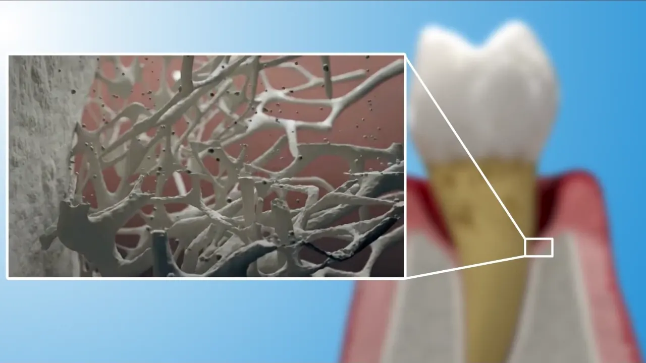
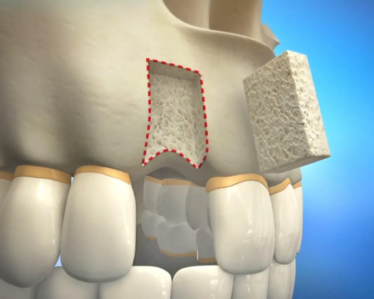
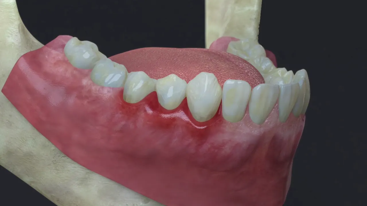
Most bone deficiencies call for a conventional bone graft beneath a flexible membrane. The mesh is for extensive defects, where bone is missing in both height and width across one or more teeth. Their causes are recognisable: advanced periodontitis that destroyed the bone around several roots before the teeth were lost; an old extraction never compensated, with the bone continuing to melt away over the years; trauma that carried off part of the ridge; more rarely, a large cyst or the removal of an implant that had lost its bone.
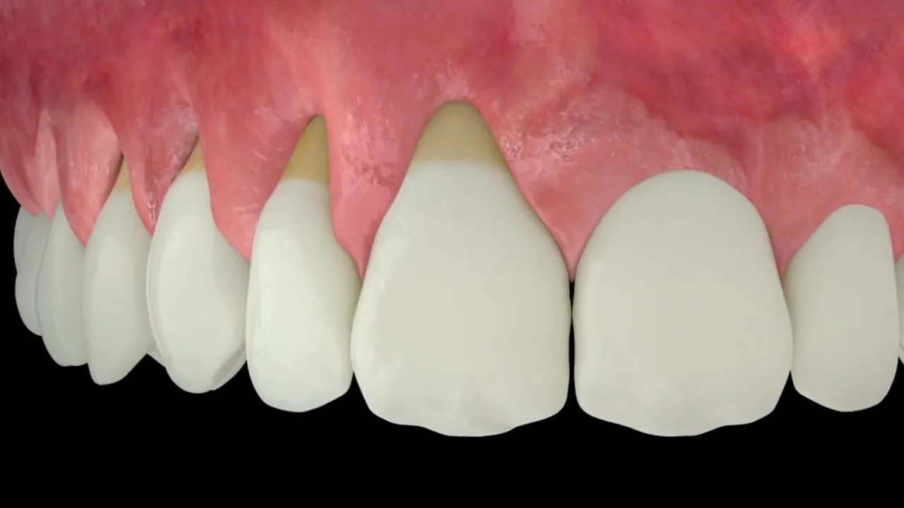
Patients, for their part, notice a sunken gum, a “hollow” ridge beneath the lip, or a removable denture that no longer stays put. Cone beam CT measures the deficit in three dimensions and shows that an implant placed in the remaining bone would be too short, too narrow or poorly aligned beneath the future crown. At that point, Dr Hazout goes through the options with you: rebuild, or do without and choose another prosthetic solution.
The role of the mesh: formwork for bone
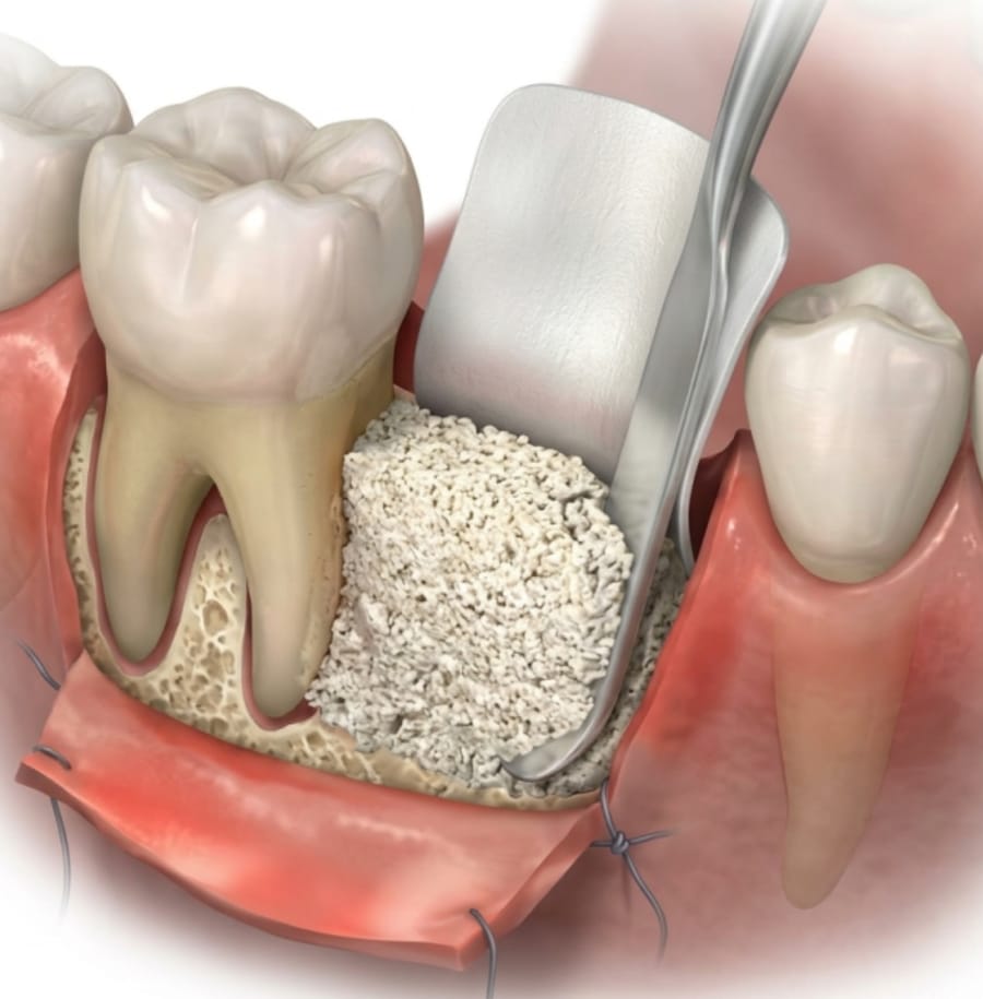
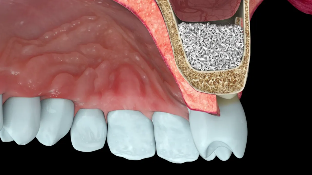
In guided bone regeneration (GBR), graft material is placed against the bone and then protected while it turns into bone. For a small defect, a flexible membrane is enough. For a large defect, especially in height, a membrane on its own collapses under pressure from the gum. The rigid mesh maintains the space: it keeps the graft in the intended shape throughout healing.
The mesh does not make bone: the graft material and the patient’s own bone see to that. It stabilises, and it protects.
The object itself
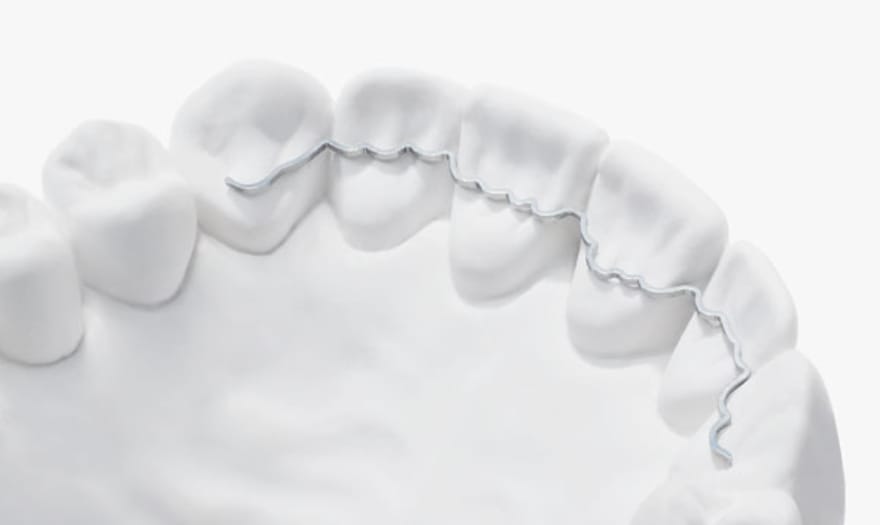
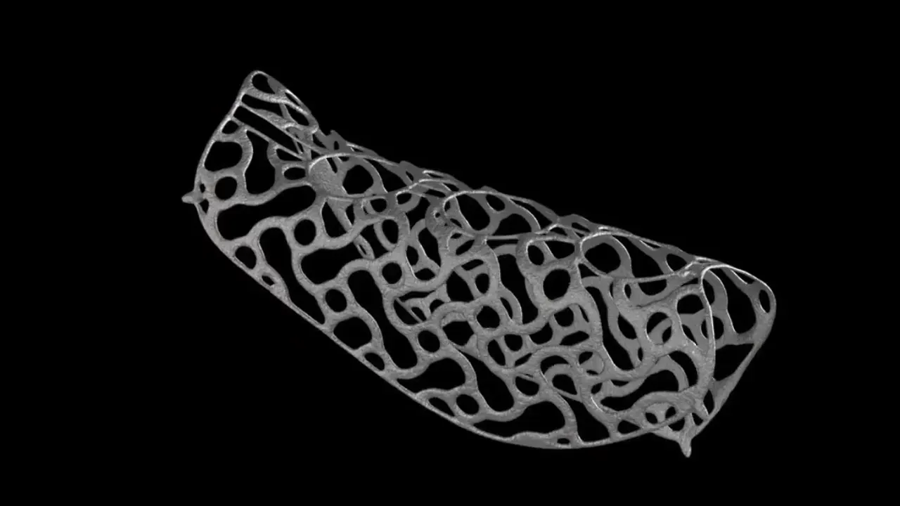
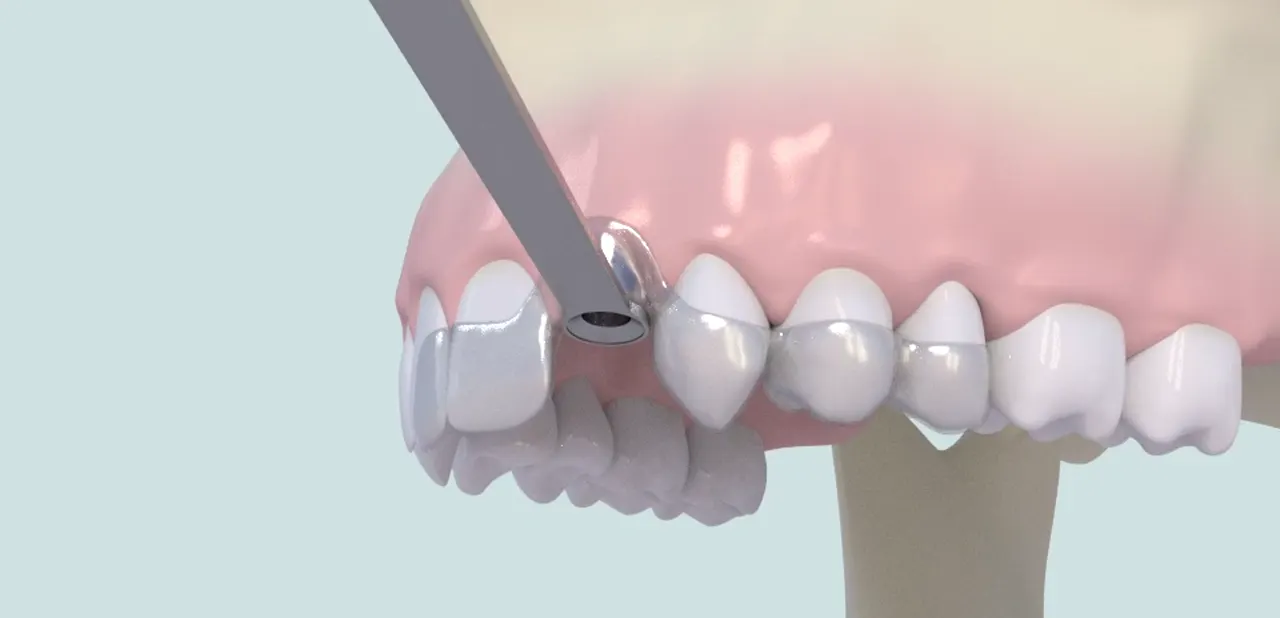
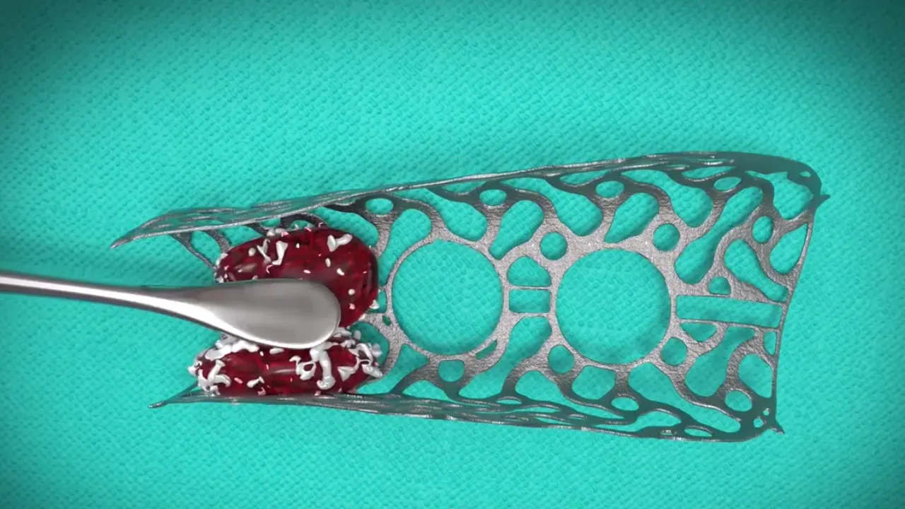
The mesh is a titanium shell a few tenths of a millimetre thick, pierced with small openings that let blood vessels reach the graft. Its shape follows the patient’s ridge closely and reproduces the volume of bone to be rebuilt. One or more small screws hold it in place.
Made to measure, from your own imaging
A conventional mesh is a standard sheet that the surgeon cuts and bends during the operation. A custom mesh is designed beforehand: from the cone beam CT (3D dental scan), the bone is reconstructed on the computer, the target volume is drawn, then the mesh that wraps around it. All that remains is to make it, by metal 3D printing.
The expected advantages: a precise fit to the bone, fewer sharp edges and a shorter operating time [1]. The manufacturing itself is the subject of the article How is a titanium mesh 3D-printed?
Planning
The mesh is not designed for its own sake, but for the implants. In the software, the future implants are first placed where the crowns call for them; the bone volume that must surround them is built next; the mesh finally follows that volume.
In the lower jaw, the path of the inferior alveolar nerve (which gives the lip and chin their sensation) is traced so that screws and implants keep their distance from it.
The grafting surgery, and the stages of treatment
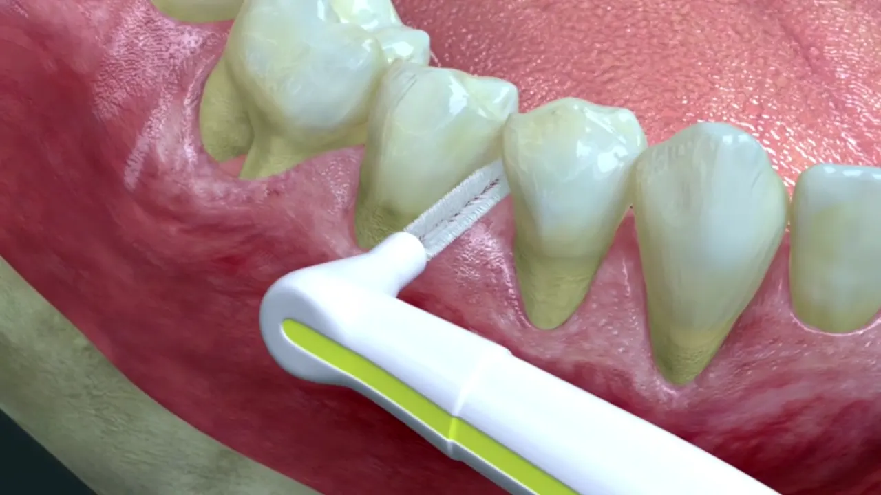
- Preparation: healthy gums, stable periodontitis, stopping smoking strongly advised.
- Grafting surgery: under local anaesthetic, in the operating room of the practice in Levallois-Perret, the gum is lifted; the mesh, packed with graft material (often a mix of the patient’s own bone and processed bovine bone), is screwed onto the ridge and covered with a membrane; the gum is sutured without tension.
- Healing: bone forms beneath the mesh, while the gum covering it is kept under watch.
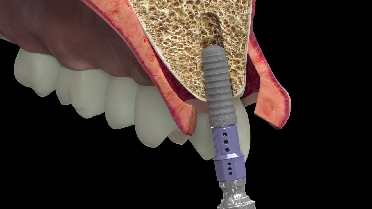
- Second surgery: the mesh is removed and the implants placed in the rebuilt bone, often during the same session.
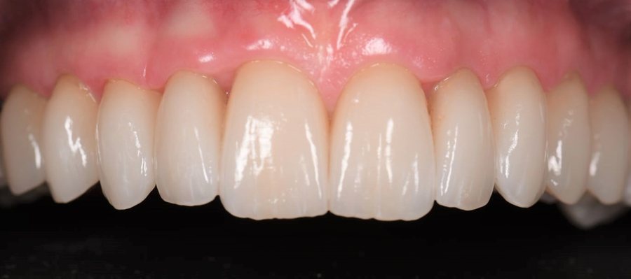
- Restoration: osseointegration secured, the crowns are fitted.
Removing the mesh
The mesh is not meant to stay: once the bone has formed, it is taken out during a second operation under local anaesthetic. The gum is lifted again, the screws removed, the mesh freed. Beneath it there is usually a thin layer of fibrous tissue, 1 to 2 mm thick, the “pseudo-periosteum”, capping the regenerated bone [4]; depending on the situation it is removed or left in place, and the underlying bone then becomes visible and measurable.
When the volume obtained allows it, the implants are placed in the same session, in the position settled at the initial planning stage. Otherwise, a small additional graft is carried out at the same time. The practical arrangements, the course of treatment and the instructions are explained to you at the practice, according to your situation.
Results and complications: what the publications say
A 2025 meta-analysis (22 studies, 608 patients) sets custom meshes against conventional ones: comparable vertical gain (5.1 mm versus 6.2 mm, difference not significant), and greater horizontal gain with the custom mesh (6.4 mm versus 3.9 mm) [1]. Most of the studies are retrospective and small.
The major complication is exposure of the mesh: the gum opens and uncovers the metal. It affects roughly one case in five with custom meshes (20.3% in the meta-analysis, against 30.9% with conventional meshes, difference not significant) [1]. Published series range from 21% [2] to 33% [3]. Exposure does not mean failure: in one series, 8 of the 11 exposed sites still integrated the graft [2]; but early exposure goes hand in hand with less bone formation [4].
In these series, survival of the implants placed afterwards reached 100% at about one year of follow-up [2, 3]; still a short follow-up.
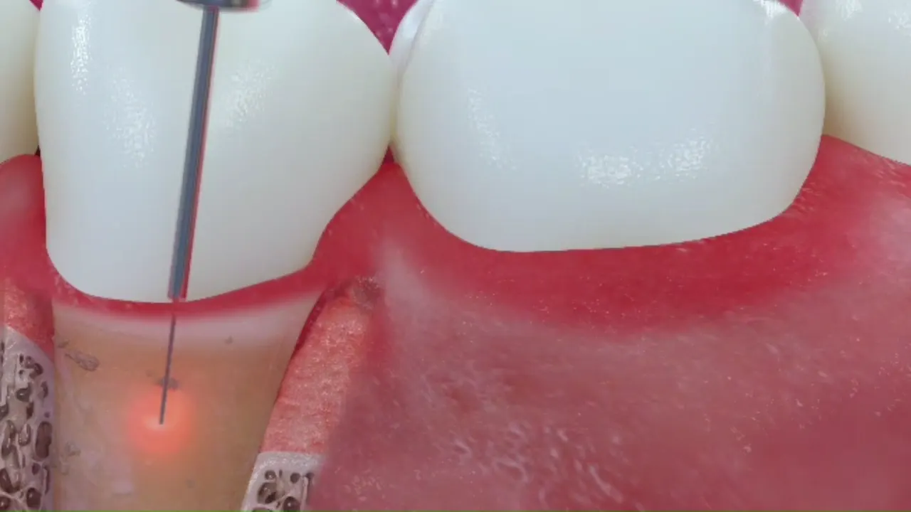
These figures come from the literature, not from a series at the practice: the animations on this page illustrate the technique as Dr Hazout performs it; they are not a record of results. Your own situation (extent of the defect, quality of the gum, smoking, general health) may place you above or below these averages; that is precisely what the assessment weighs up.
Alternatives
A mesh is only one option among those of bone regeneration: titanium-reinforced membrane, bone block or, in the lower jaw, short implants without grafting, whose five-year results are comparable to those of vertical augmentation, with fewer complications [5]. The data and limits of each are set out on the bone grafts page. The choice is discussed according to the size of the defect, the condition of your gums and your priorities.
Frequently asked questions
Does the mesh stay in the mouth for life?
No. It is removed at the second operation, once the bone has formed, at the time the implants are placed.
What happens if the mesh becomes visible?
Depending on the extent and timing of the exposure, local hygiene is reinforced with an antiseptic, or the mesh is removed earlier than planned. Exposure does not always lead to loss of the graft.
Can I be allergic to titanium?
Reactions to titanium are held to be rare. If you have a history of metal allergy, mention it at your assessment.
Scientific sources (5)
- Ragucci GM, Fernández Augè A, Tresserra Parra A, Elnayef B, Hernández-Alfaro F. Comparison between CAD/CAM titanium mesh vs. conventional titanium mesh in bone regeneration: a systematic review and meta-analysis. Int J Implant Dent. 2025;11:55. DOI
- Chiapasco M, Casentini P, Tommasato G, Dellavia C, Del Fabbro M. Customized CAD/CAM titanium meshes for the guided bone regeneration of severe alveolar ridge defects: Preliminary results of a retrospective clinical study in humans. Clin Oral Implants Res. 2021;32:498-510. DOI
- Sagheb K, Schiegnitz E, Moergel M, Walter C, Al-Nawas B, Wagner W. Clinical outcome of alveolar ridge augmentation with individualized CAD-CAM-produced titanium mesh. Int J Implant Dent. 2017;3(1):36. DOI
- Xie Y, Li S, Zhang T, Wang C, Cai X. Titanium mesh for bone augmentation in oral implantology: current application and progress. Int J Oral Sci. 2020;12:37. DOI
- Terheyden H, Meijer GJ, Raghoebar GM. Vertical bone augmentation and regular implants versus short implants in the vertically deficient posterior mandible: a systematic review and meta-analysis of randomized studies. Int J Oral Maxillofac Surg. 2021;50(9):1249-1258. DOI
Page written by Dr Victor Hazout, periodontist · updated on 7 October 2026
Patient pathway