





Placing an implant: the steps, and what guided surgery brings
An implant is placed long before the day of surgery. 3D imaging decides where it should go; the surgical guide carries that decision into the mouth. This page follows the order the patient lives through: the examination, the choice of position, the operation, fitting the tooth, and then what studies say about accuracy, lifespan and risk factors.
Contents8 sections
First of all: 3D imaging with cone beam CT
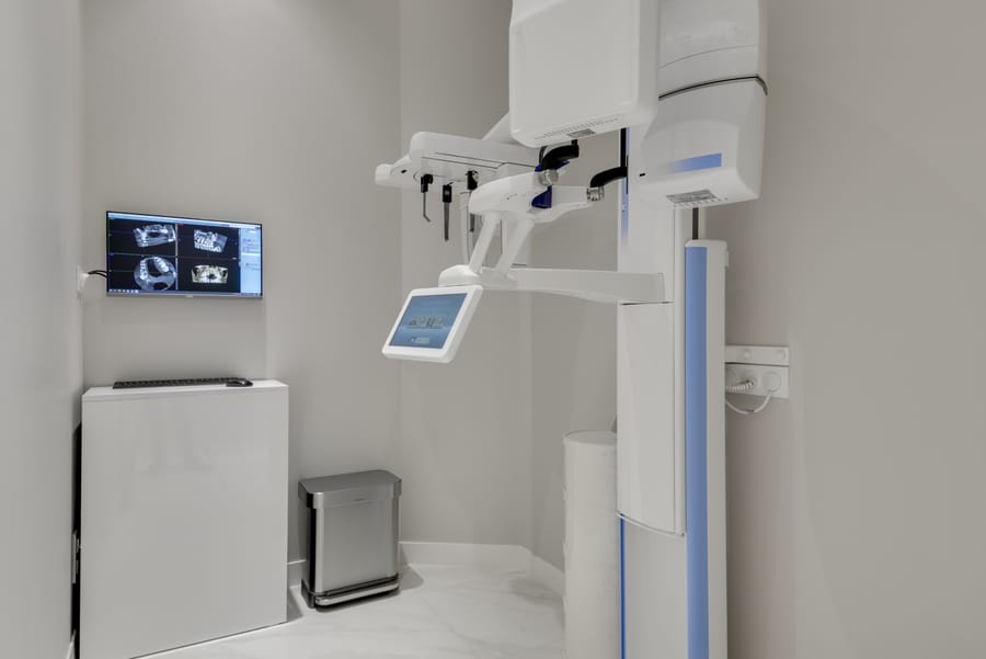
A cone beam CT is a dental scanner that renders the bone in three dimensions. It reveals the available height and width, the path of the nerve in the lower jaw and the position of the maxillary sinus. A review of recommendations judges it justified for implant diagnosis and planning, provided the field of view and the dose are adjusted to each case: depending on the machine, that dose ranges from the equivalent of 2 to 200 panoramic radiographs [1]. Metal objects (crowns, implants) and patient movement can produce artefacts that blur the image [1].
This scan goes hand in hand with an assessment of the gums and neighbouring teeth. If periodontitis is present, it is treated and re-evaluated before any placement: an implant placed in an infected mouth starts at a disadvantage.
Where should the implant go? The decision starts from the tooth to be replaced
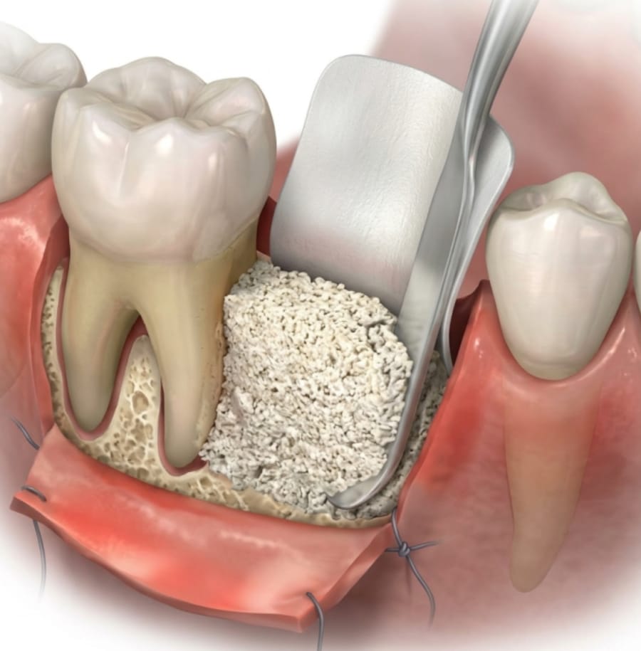
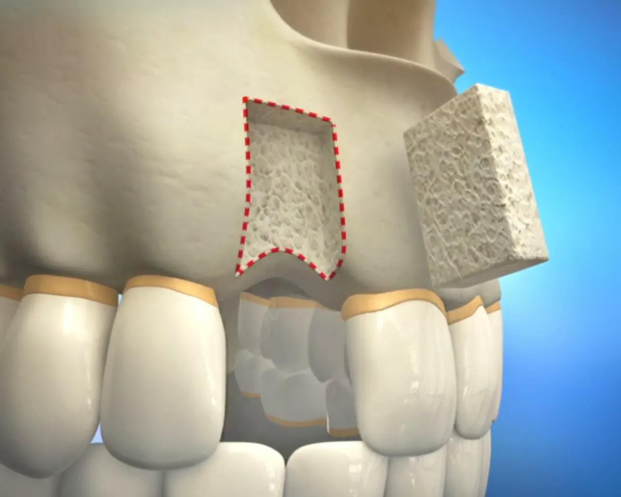
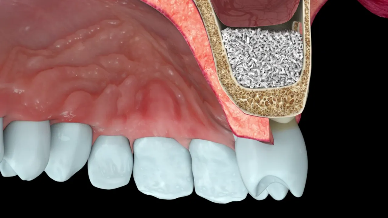
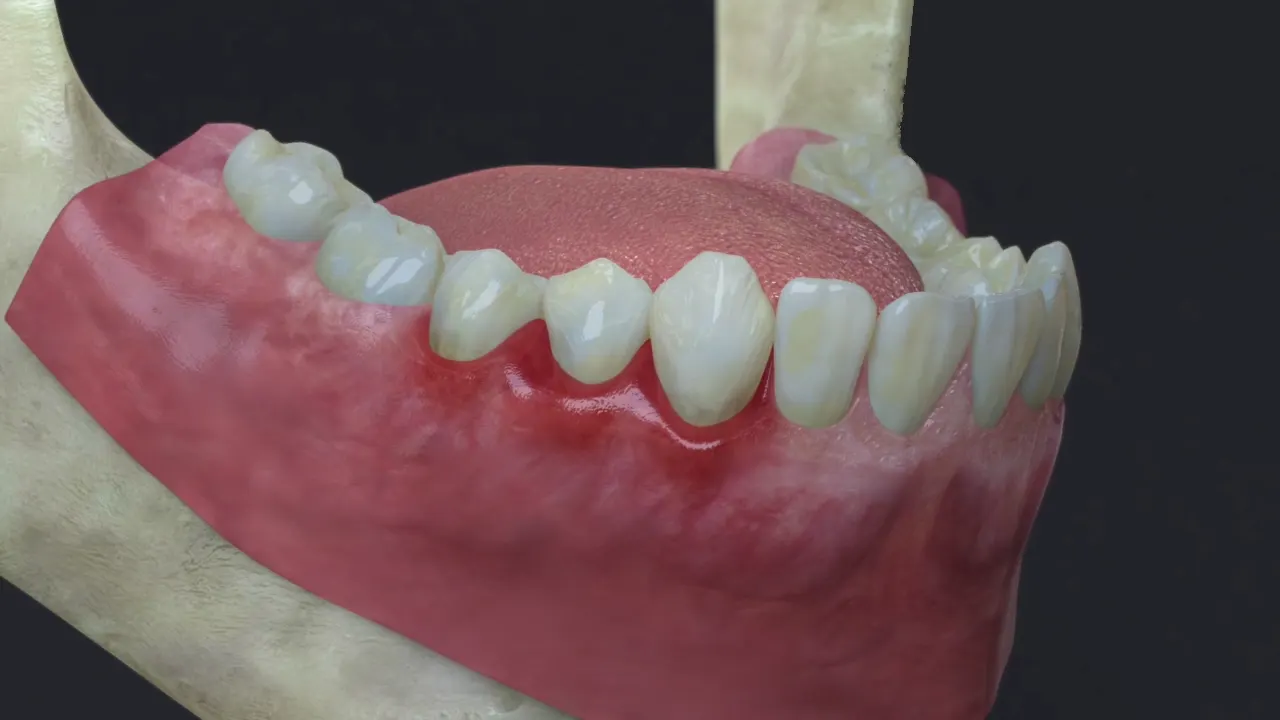
An implant is not placed where bone is most plentiful, but where the future crown needs to sit in order to chew properly and be cleaned with ease. On screen, Dr Hazout therefore designs the tooth to be replaced first, then checks that the bone can receive an implant beneath it, at a safe distance from the nerve, the sinus and the neighbouring roots. If bone is lacking at that spot, a bone graft is what gets discussed, not a retreat of the implant to a less favourable position.
The full reasoning behind this planning, what cone beam CT measures and what it does not, are developed in the article Why plan in 3D before a bone graft and an implant?
The surgical guide
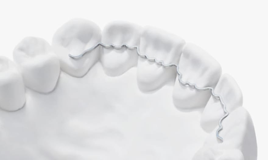
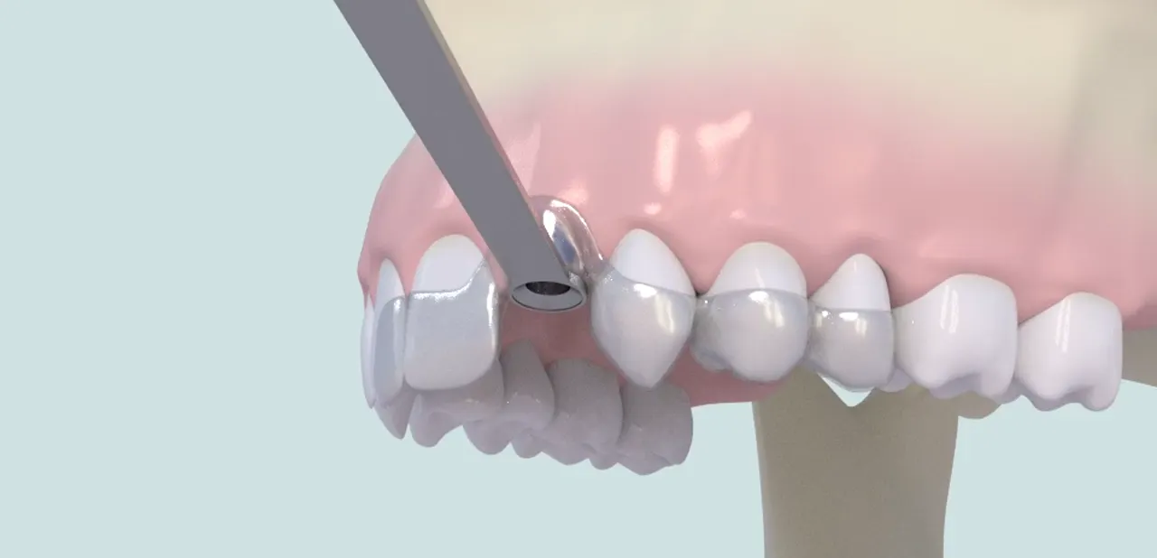
The surgical guide is a splint, usually 3D-printed in resin, that rests on the teeth or gum. Its sleeves steer the drills along the axis decided on the computer. Guidance is called fully guided when all the drilling and the insertion pass through the guide, and partially guided when only the first drill is guided.
A guide earns its place when the margin for error shrinks: an implant close to the nerve or sinus, a narrow ridge, several implants whose axes must run parallel, or placement through the gum without lifting it. For a single tooth in wide bone, well-prepared freehand placement remains a common option.
Guided or freehand surgery: how accurate?
A meta-analysis of 20 clinical studies (2,238 implants) records, with a static guide, a mean deviation of 1.2 mm at the entry point, 1.4 mm at the implant tip and 3.5° in angle compared with the plan [2]. A more recent meta-analysis sets the techniques side by side [3]:
| Technique | Angular deviation | Entry deviation | Apex deviation |
|---|---|---|---|
| Freehand | 7.5° | 1.6 mm | 2.2 mm |
| Partial guide (first drill) | 5.9° | 1.1 mm | 1.4 mm |
| Fully guided | 2.6° | 0.7 mm | 0.9 mm |
Full guidance is therefore more accurate than partial guidance, a finding confirmed by another meta-analysis [4]. But more accurate does not automatically mean better in the long run: an umbrella review of systematic reviews found no difference in implant survival or bone loss between guided and freehand surgery, and suggested that guidance mainly lowers surgical risk in complex treatments [5]. The authors recommend preserving a safety margin of at least 2 mm from sensitive structures [2, 3].
How is the implant placed?
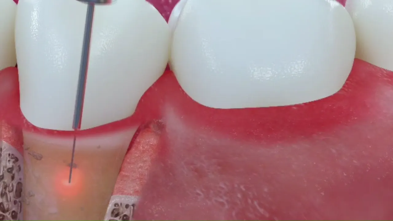
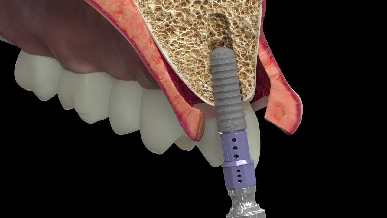
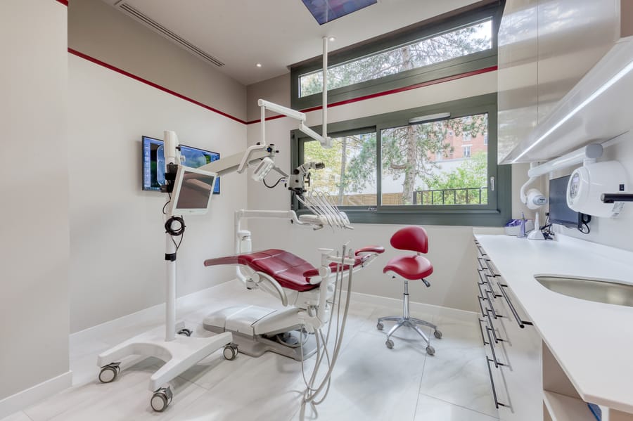
Placement takes place under local anaesthetic, in the practice’s operating room in Levallois-Perret. Depending on the case, the gum is opened to expose the bone, or the implant passes through a small opening when the guide allows it; it takes the position settled during planning. It is then either covered by the gum while healing takes place, or left flush, capped with a small healing component that shapes the gum. The practical arrangements, the course of treatment and the aftercare instructions are explained to you at the practice, according to your situation.
Healing and loading
Loading is the moment the implant receives a tooth. It can be immediate (shortly after placement), early, or conventional (once osseointegration is achieved). A Cochrane review of 26 randomised trials found no clinically important difference in failure between these options, with very low certainty of evidence [6]. The choice depends on how stable the implant is at placement, on bone quality and on whether a graft was needed.
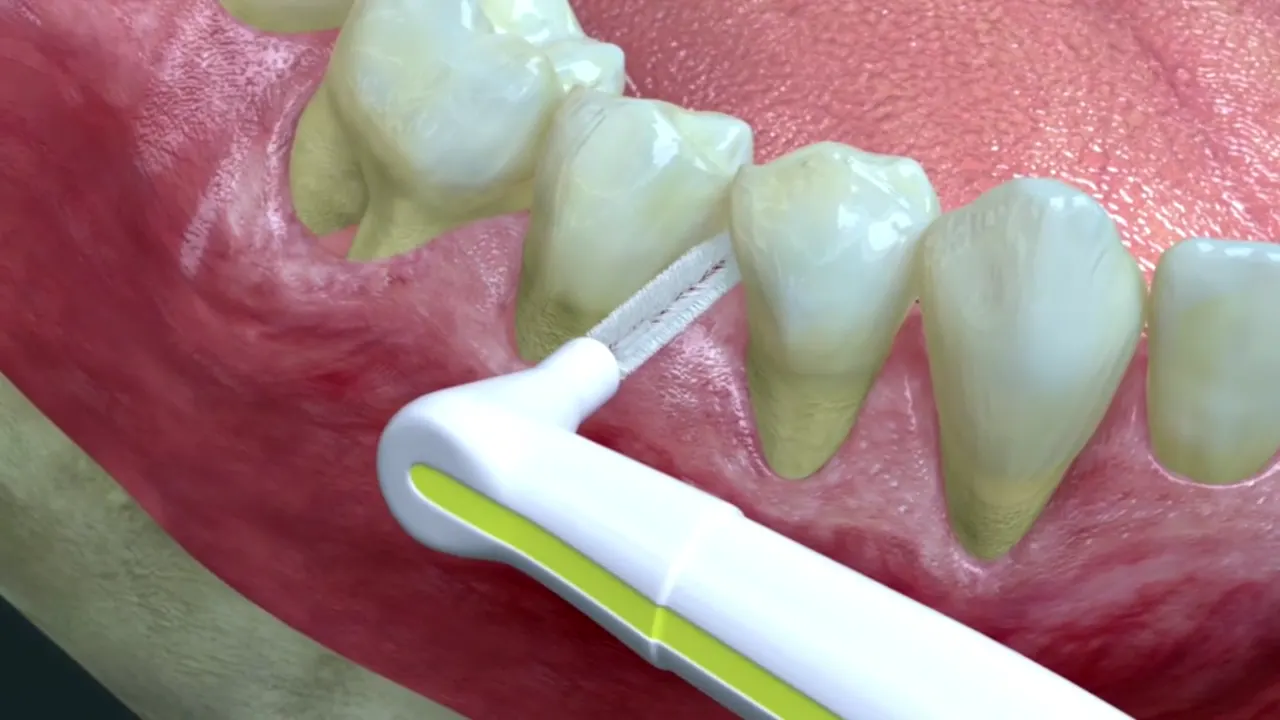
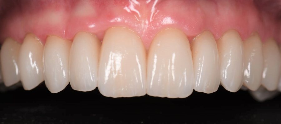
Once osseointegration is achieved, an impression is taken and the final crown, screwed or cemented onto an abutment, is fitted. Its design shapes what follows: a crown that lets interdental brushes through can be cleaned, whereas a crown that is too bulky at the neck traps plaque.
How long does an implant last?
A meta-analysis estimates ten-year survival at 96.4%, and 93.2% in a more conservative sensitivity analysis [7]. These figures are averages: your personal risk rests on factors that can, in part, be changed.
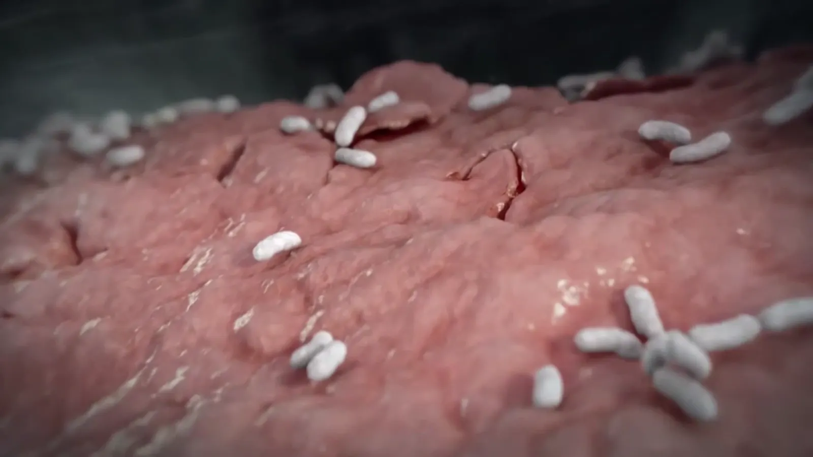
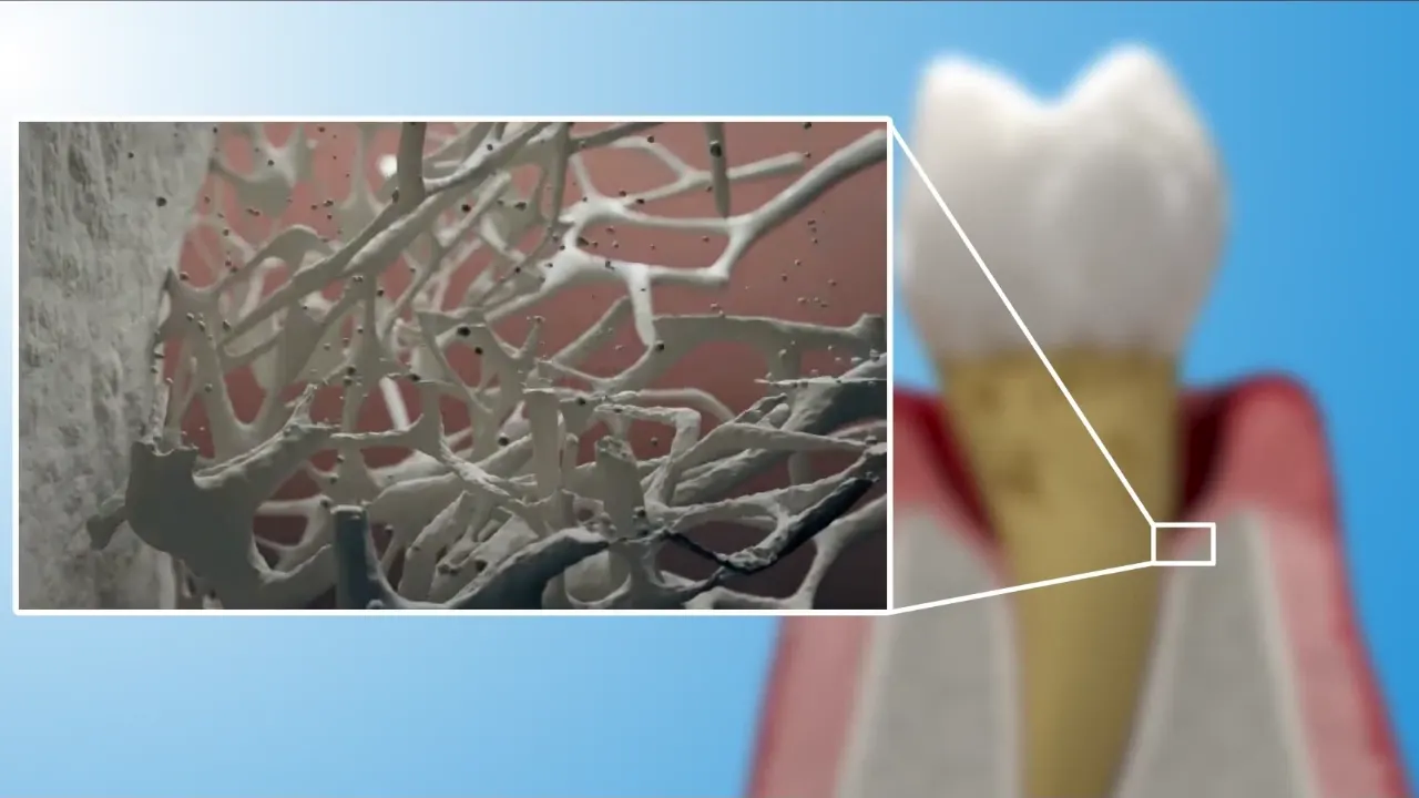
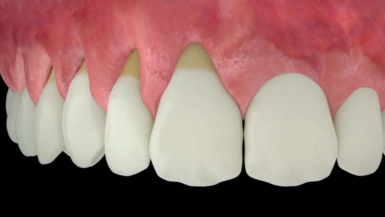
The main long-term threat is not mechanical but infectious: peri-implantitis, inflammation of the tissues around the implant with loss of supporting bone. It is prevented by appropriate daily cleaning and by maintenance visits whose frequency follows your level of risk. The article Gum care around implants details these habits.
Contraindications and risk factors
- Smoking. A systematic review of 44 studies links smoking with a higher risk of implant failure and an average of 0.64 mm of additional bone loss; it is not an absolute contraindication, but support to stop smoking should form part of the treatment plan [8].
- Poorly controlled diabetes. Poorly controlled diabetes is associated with more peri-implantitis and, in the long term, more implant loss; when it is well controlled, results match those of people without diabetes [9].
- History of periodontitis. It is associated with more peri-implantitis and slightly lower survival [10]. A review prepared for the 2017 World Workshop on the Classification of Periodontal and Peri-Implant Diseases lists a history of periodontitis, poor plaque control and lack of regular maintenance among the risk factors for peri-implantitis backed by strong evidence [11].
- Situations assessed case by case: some bone medications (antiresorptives), head and neck radiotherapy, or an unstable general condition. These are discussed with your doctor.
Frequently asked questions
Is guided surgery essential?
No. It sharpens positional accuracy, which is valuable near the nerve or sinus, in narrow bone or with several implants. For a simple case, well-planned freehand placement gives, according to the available reviews, comparable long-term results.
Why a cone beam CT rather than a panoramic X-ray?
A panoramic X-ray is a two-dimensional image that does not show bone width. Cone beam CT measures it in three dimensions. Because its dose is higher, the area scanned is limited to what is strictly needed.
Can I have a tooth on the same day?
Sometimes, if the implant is very stable at placement and the bone is of good quality. Otherwise, a removable or bonded temporary tooth takes over during healing.
Should I stop smoking before an implant?
It is strongly advised: smoking is associated with more failures and more bone loss around implants. Support to stop can be offered to you.
Scientific sources (11)
- Jacobs R, Salmon B, Codari M, Hassan B, Bornstein MM. Cone beam computed tomography in implant dentistry: recommendations for clinical use. BMC Oral Health. 2018;18:88. DOI
- Tahmaseb A, Wu V, Wismeijer D, Coucke W, Evans C. The accuracy of static computer-aided implant surgery: A systematic review and meta-analysis. Clin Oral Implants Res. 2018;29 Suppl 16:416-435. PubMed
- Werny JG, Frank K, Fan S, Sagheb K, Al-Nawas B, Narh CT, Schiegnitz E. Freehand vs. computer-aided implant surgery: a systematic review and meta-analysis—part 1: accuracy of planned and placed implant position. Int J Implant Dent. 2025;11:35. DOI
- Khaohoen A, Powcharoen W, Sornsuwan T, Chaijareenont P, Rungsiyakull C, Rungsiyakull P. Accuracy of implant placement with computer-aided static, dynamic, and robot-assisted surgery: a systematic review and meta-analysis of clinical trials. BMC Oral Health. 2024;24:359. DOI
- Tomar S, Chaudhary P, Ganguly A. Comparing the clinical outcomes of guided and freehand dental implant surgery: An umbrella review of systematic reviews and meta-analyses. J Prosthet Dent. 2026;135(5):e53-e59. DOI
- Esposito M, Grusovin MG, Maghaireh H, Worthington HV. Interventions for replacing missing teeth: different times for loading dental implants. Cochrane Database Syst Rev. 2013;CD003878. DOI
- Howe MS, Keys W, Richards D. Long-term (10-year) dental implant survival: A systematic review and sensitivity meta-analysis. J Dent. 2019;84:9-21. DOI
- Calciolari E, Corbella S, Dourou M, Ercal P, Donos N. Tobacco smoking and smoke-free products as risk factors for dental implants: A systematic review. Clin Oral Implants Res. 2026;37(3):262-286. DOI
- Wagner J, Spille JH, Wiltfang J, Naujokat H. Systematic review on diabetes mellitus and dental implants: an update. Int J Implant Dent. 2022;8:1. DOI
- Marty L, Hoornaert A, Enkel B, Penhoat A, Colat-Parros J, Soueidan A, Jordana F. Implant Health in Treated Periodontitis Patients: A Systematic Review and Meta-Analysis. Dent J (Basel). 2024;12(8):240. DOI
- Schwarz F, Derks J, Monje A, Wang HL. Peri-implantitis. J Periodontol. 2018;89 Suppl 1:S267-S290. DOI
Page written by Dr Victor Hazout, periodontist · updated on 11 October 2026
Patient pathway